This glossary will be beneficial for me to become more comfortable in this field.
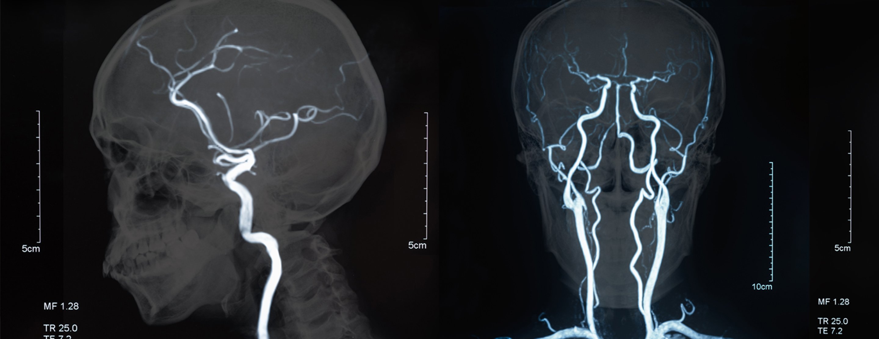
angiography
noun
A diagnostic imaging procedure that uses X-rays and a special contrast dye to visualize the inside of blood vessels and organs, particularly the heart, brain, and lungs.
Example: There was associated chest pain but this was a constant tightness rather than a sharp pleuritic pain as seen with a PE. Investigations for suspected PE: Presence of D-Dimer in blood - no specific for PE but if absent excludes PE as a differential V-Q perfusion scan - to assess ventilation perfusion mismatch CT pulmonary ANGIOGRAPHY - to assess blood flow throughout lungs Pneumothorax - Pneumothorax can occur spontaneously or as a result of trauma.
en: angiographie
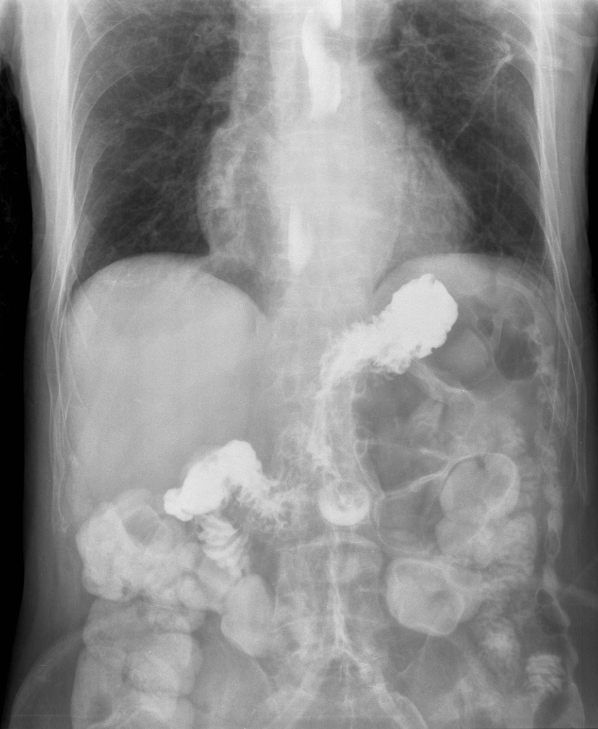
barium x-ray
noun
A diagnostic imaging test that uses a chalky, liquid contrast medium (barium sulfate) and fluoroscopy (real-time X-ray) to create detailed images of the digestive tract.
Example: The patient had a barium X-ray to help the doctor see the shape of the esophagus and detect any swallowing problems.
en: radiographie au baryum
bone densitometry
noun
A non-invasive, low-dose X-ray procedure that measures calcium and mineral levels in bones.
Example: The doctor ordered a bone densitometry test to measure the patient’s bone strength and check for signs of osteoporosis.
en: densitométrie osseuse
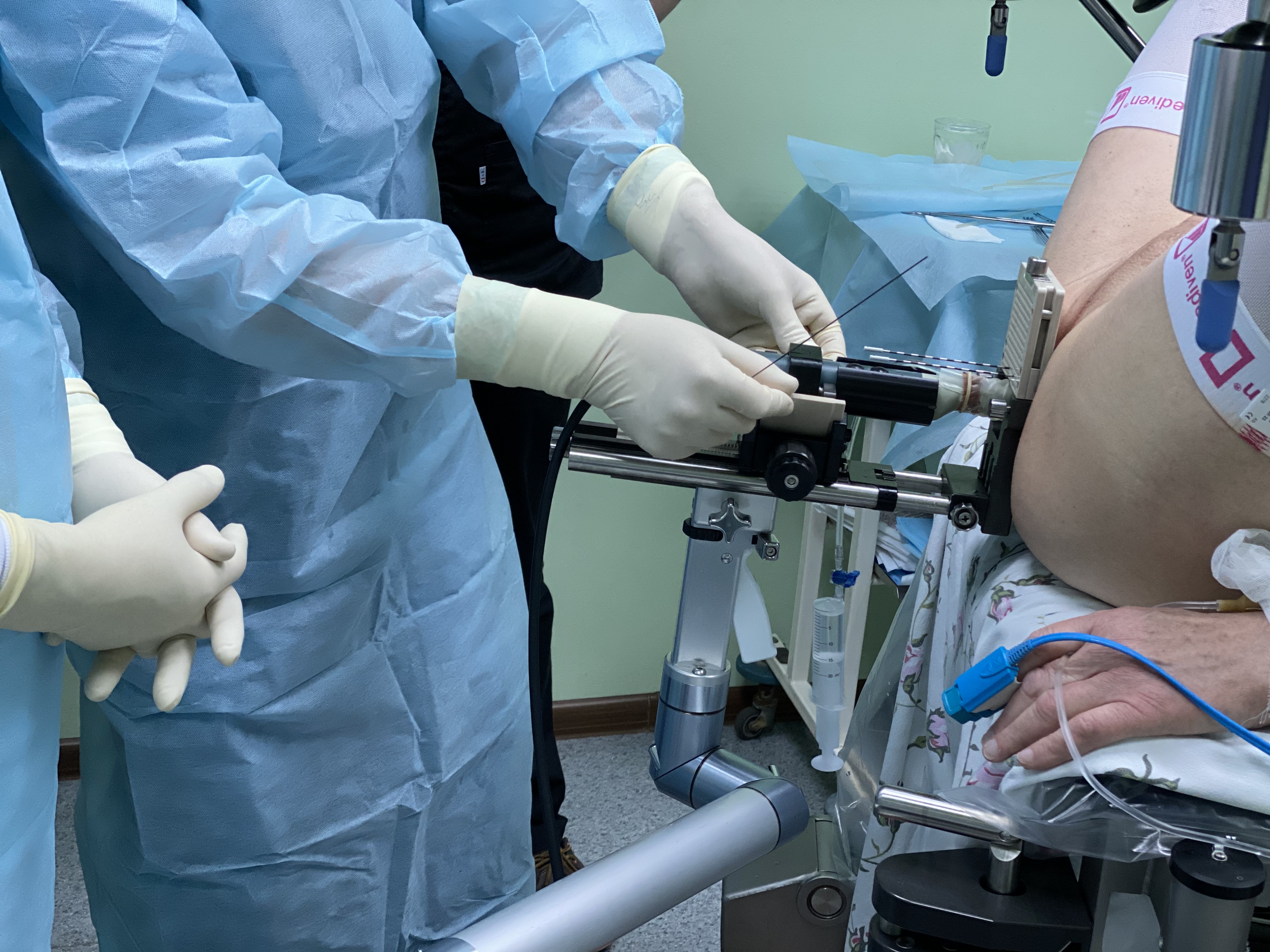
brachytherapy
noun
A type of internal radiation therapy used to treat cancer by placing radioactive sources—such as seeds, ribbons, or capsules—directly into or immediately adjacent to a tumor.
Example: The doctor recommended brachytherapy because it allows radiation to be delivered directly to the tumor while minimizing damage to nearby healthy tissue.
en: brachythérapie
bucky diaphragm
noun
A radiology device featuring a moving grid placed between the patient and the x-ray film/detector to reduce scattered radiation.
Example: The radiologic technologist adjusted the bucky diaphragm to limit the x-ray beam and improve image quality by reducing unnecessary radiation exposure.
en: diaphragme bucky
computed tomography
noun
A noninvasive, painless medical imaging procedure that uses rotating X-rays and computer technology to produce detailed, cross-sectional, 3D images of bones, blood vessels, and soft tissues.
Example: Future directions for detection of coronary reperfusion after thrombolytic therapy Coronary echocardiography, Magnetic resonance imaging (MRI), Cardiac COMPUTED TOMOGRAPHY scanner (CT) and transthoracic Doppler echocardiography (TTDE) are just some of the emerging techniques used by clinicians to accurately evaluate IRA patency, providing information on coronary reperfusion.
en: tomodensitométrie
computerized axial tomography scan
noun
A noninvasive medical imaging procedure that uses rotating X-rays and computer technology to produce detailed, cross-sectional, 3D images of soft tissues, bones, and blood vessels.
Example: After the accident, the patient underwent a computerized axial tomography scan to help doctors detect any internal injuries or bleeding.
en: tomodensitométrie axiale
contraindication
noun
A condition or circumstance that suggests or indicates that a particular technique or drug should not be used in the case in question.
Example: A severe allergy to contrast dye is considered a contraindication for certain imaging procedures.
en: contre-indication
dosimeter
noun
A small, portable device worn on the body to measure and monitor an individual's accumulated exposure to ionizing radiation (such as X-rays, gamma rays, or beta radiation) over time.
Example: The radiologic technologist wears a dosimeter to monitor the amount of radiation exposure received during work hours.
en: dosimètre
endoscopy
noun
A minimally invasive medical procedure used to visualize, diagnose, and treat conditions inside the body using an endoscope.
Example: Investigations Full blood count Liver function tests Urea and electrolytes ENDOSCOPY - Upper GI tract ENDOSCOPY showed the presence of oesophageal varices, although they were not bleeding.
en: endoscopie
femoral neck
noun
He flattened, pyramidal region of bone connecting the femoral head (the ball of the hip joint) to the femoral shaft.
Example: The X-ray revealed a fracture in the femoral neck, which explained the patient’s hip pain after the fall.
en: col du fémur
fluoroscopy
noun
A medical imaging technique that uses continuous X-ray beams to produce real-time, "movie-like" video footage of internal body structures in motion.
Example: Fluoroscopy allows doctors to observe real-time movement inside the body, such as swallowing during a barium study.
en: fluoroscopie
gallbladder
noun
A small, pear-shaped organ located in the upper right abdomen beneath the liver, crucial for storing and concentrating bile produced by the liver.
Example: GALLBLADDER is dilated, no stones, common bile duct appears normal.
en: vésicule biliaire
malignant
adverb
(of a disease) very virulent or infectious.
Example: The biopsy confirmed that the tumor was malignant and required immediate treatment.
en: maligne
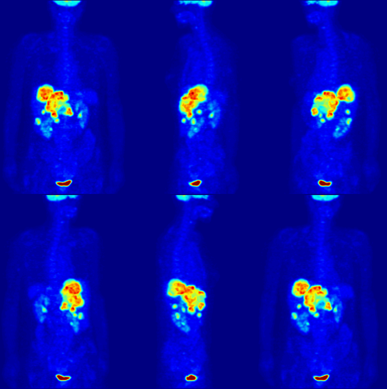
positron emission tomography scan
noun
A specialized nuclear medicine imaging technique that detects diseases by visualizing the metabolic activity of tissues and organs.
Example: The doctor recommended a positron emission tomography scan to check how the patient’s brain was functioning and to look for signs of disease.