20 Learning the basics of the machines in the department and how the images are made. terms
I'm entering my third year in this program and it's been a very interesting experience. I made this glossary to make a brief summary of how our machines work and for people who might be interested in that subject. X-rays have been used for decades in the medical field to diagnose many illnesses and help people in need for treatment and the technology keeps evolving throughout the years making it more efficient and performing.
angiography
noun
Examination by X-ray of blood or lymph vessels, carried out after introduction of a radiopaque substance.
Example: Coronary ANGIOGRAPHY is considered to be the gold standard for the detection of reperfusion as it provides an accurate visualisation of the occluded IRA and predicts TIMI flow.
fr: angiographie
anode
noun
The positively charged electrode by which the electrons leave a device.
Example: some of the photoelectrons can pass from the cathode to the ANODE, and so a small current is created.
fr: anode
barium enema
Noun
A barium enema is an X-ray exam that can detect changes or abnormalities in the large intestine (colon). The procedure is also called a colon X-ray.
Example: Imaging - Small bowel enema-(introduction of barium directly into small intestine via tube), and also air-contrast BARIUM ENEMA are the best techniques for barium imaging.
fr: Lavement baryté
cathode
noun
The negatively charged electrode by which electrons enter an electrical device.
Example: X-rays were discovered over a century ago by certain circumstances involving CATHODE-rays (streams of electrons) hitting various materials.
fr: cathode
central ray
Noun
The central ray is a simplified way of indicating the direction in which an X-ray beam travels.
Example: During an exam, I'll need to put the central ray at the precise area to make a good image without missing any structure.
fr: Rayon central
compensating filter
noun
An X-ray filter (or compensating filter) is a device placed in front of an X-ray source in order to reduce the intensity of particular wavelengths from its spectrum and selectively alter the distribution of X-ray wavelengths within a given beam before reaching the image receptor.
Example: To reduce the dose to the patient and create more detail in the image, we use a compensating filter.
fr: Filtre compensateur
coronarography
Noun
Coronary angiography is a procedure that uses a special dye (contrast material) and x-rays to see how blood flows through the arteries in your heart.
Example: You'll have to go through an coronarography if your blood vessels are blocked.
fr: Coronarographie
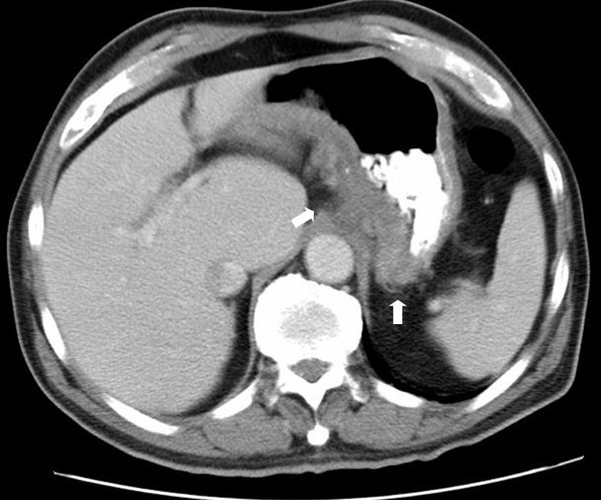
CT scan
Noun
An X-ray image made using a form of tomography in which a computer controls the motion of the X-ray source and detectors, processes the data, and produces the image.
Example: I found it very surprising that, in a patient where staging and investigation into the extent of his cancer was critical not only to his management but also for himself and his family, that he had to wait almost 2 weeks for a CT SCAN.
fr: Tomodensitométrie
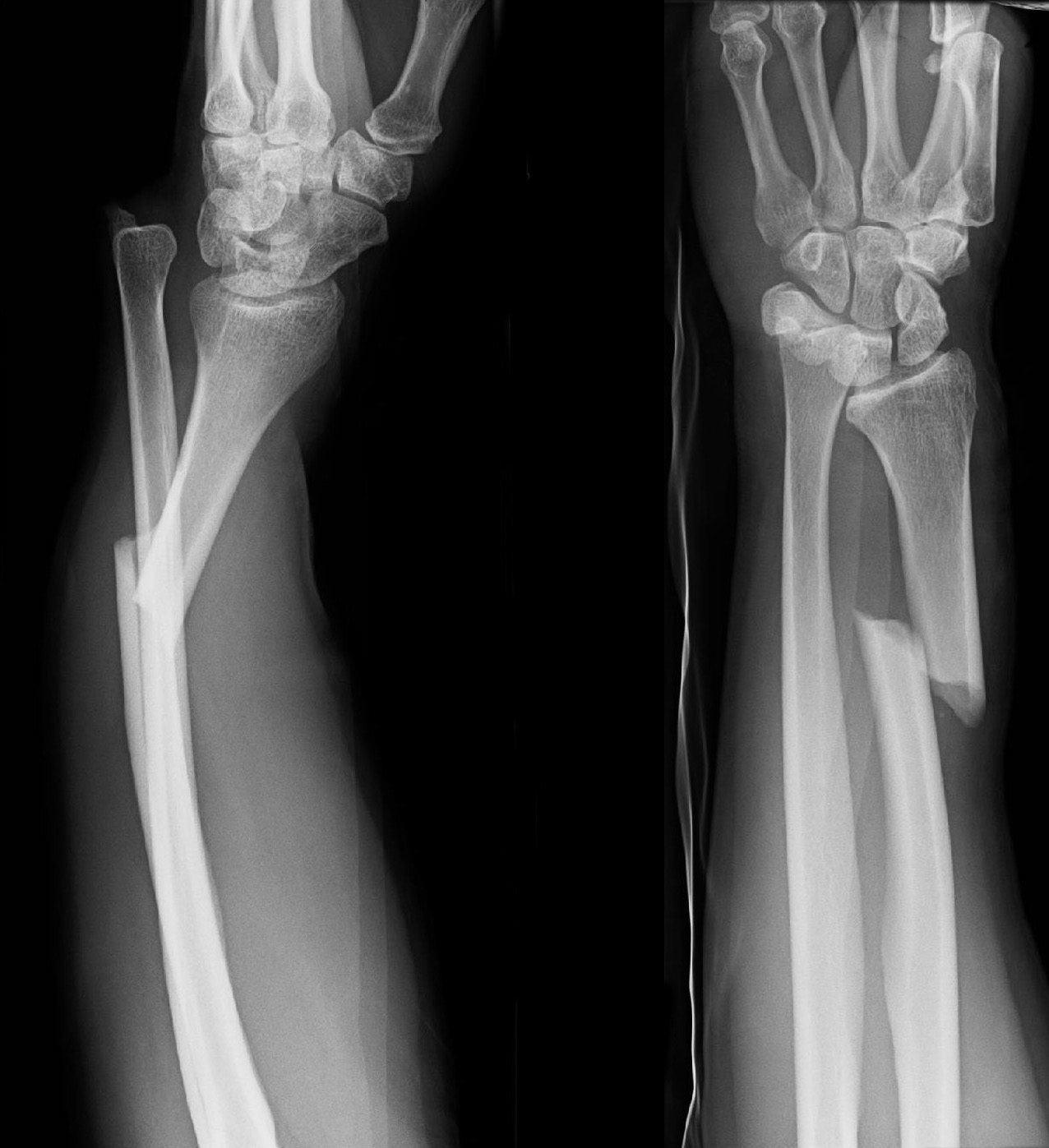
fracture
Noun
The act or process of breaking or the state of being broken.
Example: A decrease in bone cross-sectional area, reducing its ability to resist stresses and to resist fracture.
fr: Fracture
Kilovoltage peak (kVp)
noun
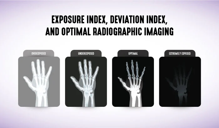
The kVp is the difference in potential or energy applied to the X-ray tube.
Example: The higher the kilovoltage peak is, the more the protons will have energy to go through tissues and bones.
fr: Kilovoltage
lead apron
Noun
A lead apron is a protective garment worn by medical personnel to shield themselves from radiation exposure.
Example: I need to wear a lead apron if I do an x-ray in a patient's room.
fr: Tablier plombé
Magnetic resonance imaging
Noun
A form of medical imaging that measures the response of the atomic nuclei of body tissues to high-frequency radio waves when placed in a strong magnetic field, and that produces images of the internal organs.
Example: X-rays are not used in magnetic resonance imaging compared to other machines.
fr: Imagerie en résonance magnétique
mammography
noun
A technique using x-rays to diagnose and locate tumors of the breasts.
Example: For example, a new study indicates that MRI (71%) is more sensitive than MAMMOGRAPHY (40%) for detecting cancers in women with inherited susceptibility.
fr: mammographie
Milliampere-seconds
noun
Milliampere-seconds, also more commonly known as mAs, is a measure of radiation produced (milliamperage) over a set amount of time (seconds) via an x-ray tube
Example: In radiology we use a different milliampere-second (mAs) with each structure to get a visible and clear image.
fr: Miliampère par seconde
noise (in radiology)
Noun
It appears as an irregular granular pattern in all images and degrades image information.
Example: This particular image is lacking in detail and quality because of the noise.
fr: Bruit radiologique
Pathology
noun
The science of the causes and effects of diseases.
Example: Seeing some type of abnormal mass or problem within a person's body in our image is considered a pathology.
fr: Pathologie
Proton
noun
a stable subatomic particle occurring in all atomic nuclei, with a positive electric charge equal in magnitude to that of an electron, but of opposite sign.
Example: Protons are used in the processing of creating an image for diagnostic imaging.
fr: Proton
Radioscopy
noun
Examination of the inner structure of opaque objects using X rays or other penetrating radiation.
Example: While it is very similar to radiography, radioscopy makes you see the image like a live video instead of a picture.
fr: Radioscopie
X-ray
noun
An electromagnetic wave of high energy and very short wavelength, which is able to pass through many materials opaque to light.
Example: I feel a great amount of pain in my lower rib cage area. I might need to go to the hospital to get an x-ray.
fr: radiographie
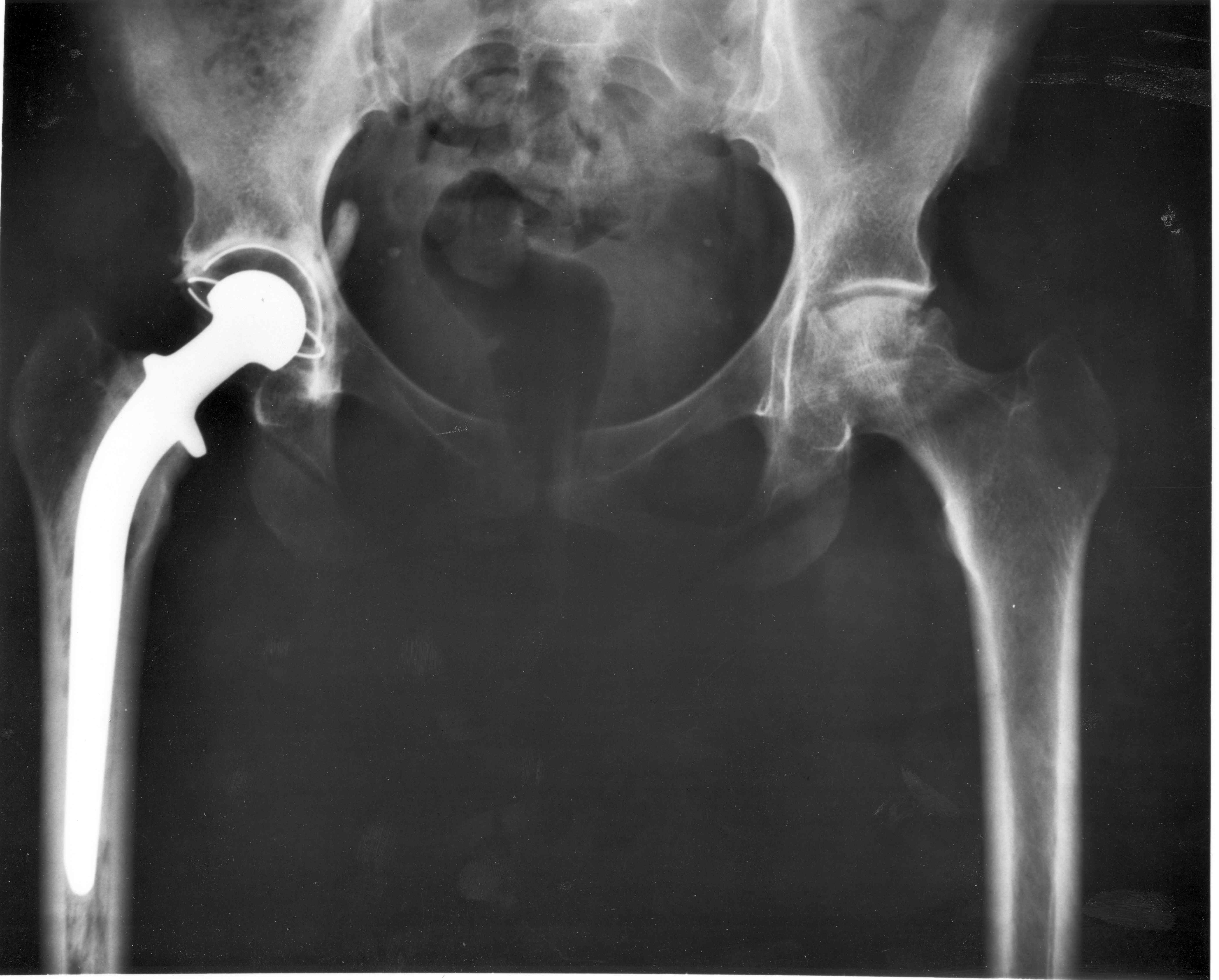
x-ray artefacts
Noun
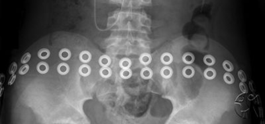
An X-ray artifact is a flaw in the image quality that can obscure what you were attempting image. It can also cause a misdiagnosis as an artifact may be mistaken for a foreign body.
Example: Any object that can be removed that isn't considered as a part of the body is an x-ray artefact.